

The following is a collection of scanning electron micrographs taken as a part of ongoing research initiatives at SMS. They highlight patterns that are being developed for active projects and internal research as well as relevant patterns found in nature.

Molds
Metal molds created using our seamless nanopatterning process, nanocoining.

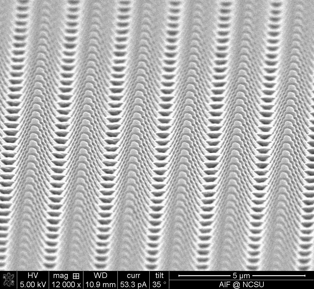
Ripples in the diamond die were replicated into the electroformed copper mold

Ripples in the diamond die were also replicated into the high phosphorous nickel mold
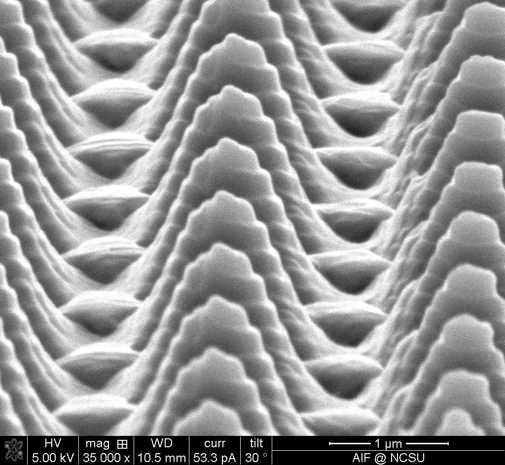
Material flow resulting from the nanocoining indenting process

Close up of material flow due to indenting


Polymer Replicas
Following the fabrication of a master mold, the pattern can be transferred using nanofabrication techniques including thermal embossing and nanoimprint lithography.
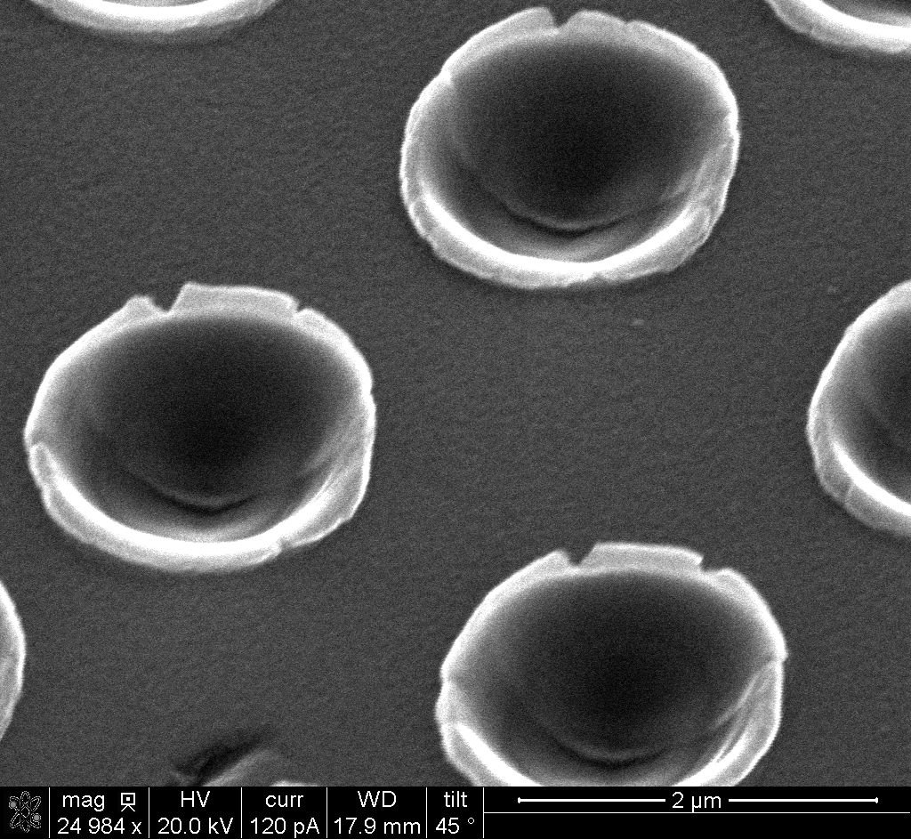
Nanoimprint lithography (NIL) with a mold created with nanocoining at SMS was used to pattern this resist. Subsequent etching steps transferred this pattern into a metal layer to create a metal-mesh film for plasmonic IR absorbers.


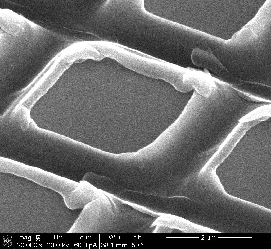
Replica Flaws
Wrinkles and bubbles in replicas


A bubble between a UV-cured grid pattern and its polycarbonate backing


Interwoven Indents
Intentional tiling errors during the nanocoining process create patterns consisting of differently sized features.

A micropattern replicated into a photopolymer using a flexible shim and a batch NIL process

A pattern replicated using one of our seamless sleeves and a UV Roll-to-Roll NIL process

Nature
Many engineered nanopatterns find inspiration from nature. The biomimetic effects include hydrophobicity and structural color.
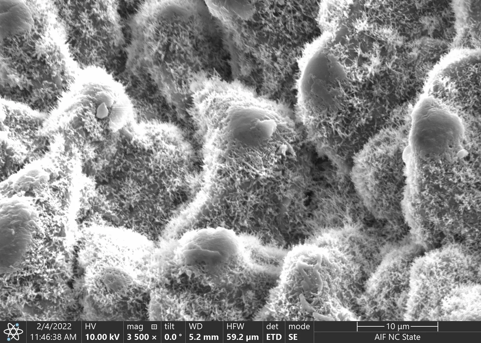


Intricate hierarchical patterns make lotus leaves self-cleaning and hydrophobic.

The microscopic tree-like structures give Morpho butterfly wings their vibrant blue color through a phenomenon known as structural color This image resembles a forest with tree-covered hills.
These images were taken by Lauren Micklow, Nicky Cates, and Brenna Tryon at the North Carolina State University Analytical Instrumentation Facility using an FEI Quanta 3D DualBeam SEM/FIB or a Helios 5 Hydra DualBeam SEM/pFIB.